Arteries In Neck Labeled : nemfrog - Arteries of the neck. Applied Anatomy: Designed... / 1st innominate divides into right subclavian and right common carotid.
Arteries In Neck Labeled : nemfrog - Arteries of the neck. Applied Anatomy: Designed... / 1st innominate divides into right subclavian and right common carotid.. 1st innominate divides into right subclavian and right common carotid. Find this pin and more on anatomy and pysiology block 2by tonna brinson. The principal arteries are the carotid and subclavian arteries. There are 4 main arteries in your neck; Branches of arch of aorta.
The first branch of the thyrocervical trunk is the inferior thyroid artery. Labeled diagram of the arteries of the head and neck. The internal carotid artery (latin: Branches of arch of aorta. Bodytomy provides a labeled iliac artery diagram to help you understand the anatomy and function of the common iliac.
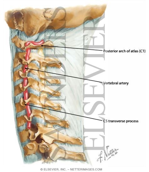
Vertebral Artery, Neck from www.netterimages.com The internal carotid artery (latin: Label the arteries of the neck in the ct angiogram. Simple labelled illustration depicting the general pathways for the major arteries of the head and neck. The external carotid artery has eight named branches distributed to the head and neck. Labeled diagram of the arteries of the head and neck. After denudation, all injured carotids of wt mice showed a higher mean. A blockage in one of the carotid arteries can be cleared either by endarterectomy or carotid angioplasty. From this trunk, several vessels arise, which go on to supply the neck.
Transverse cervical artery is labeled, branching from the thyrocervical_trunk.
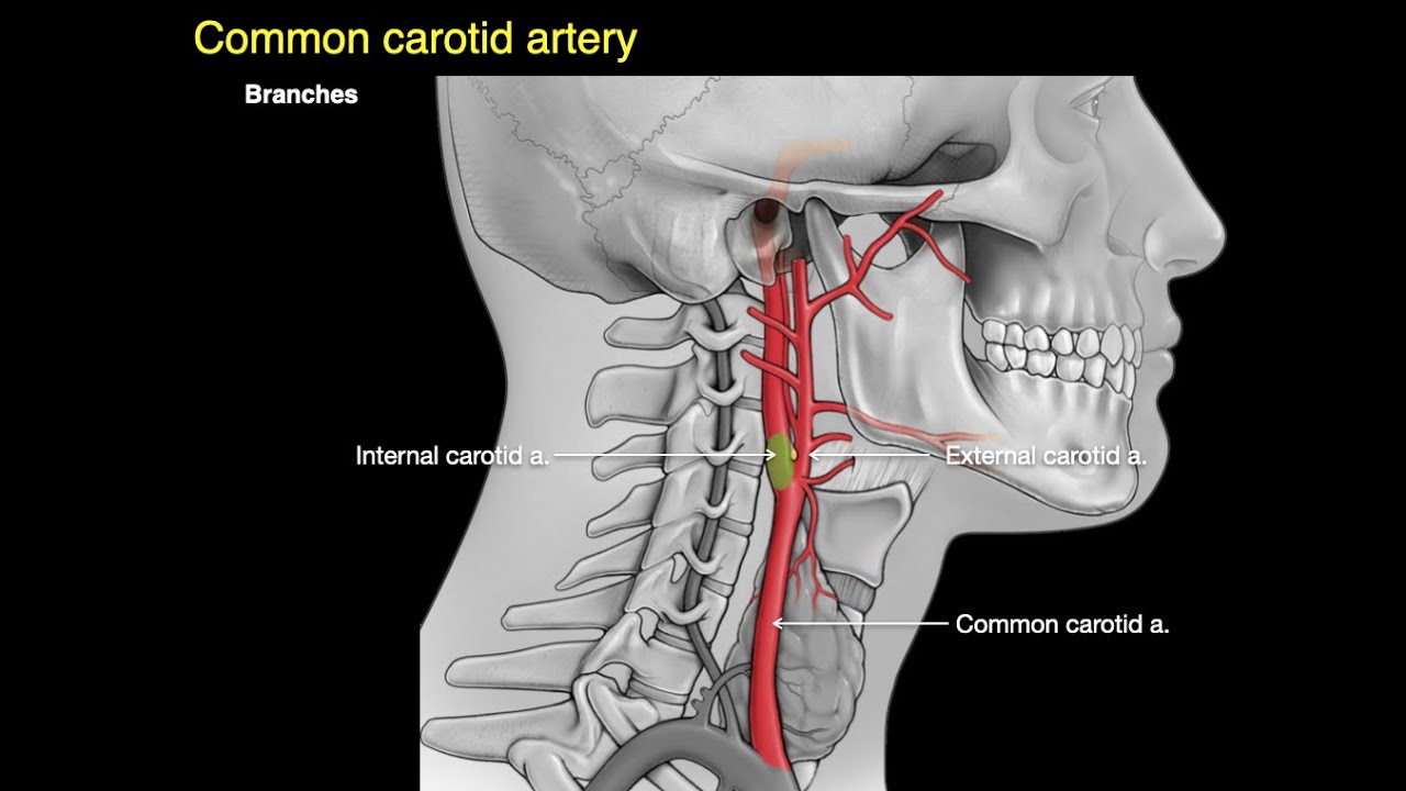
Variable in extent, the platysma typically spans the space between the superior margins of pectoralis. Terms in this set (32). Illustration showing arteries in the head noted at right are anterior cerebral artery middle cerebral artery internal carotid artery and common carotid artery labeled at left is posterior cerebral arteries in the neck the carotid arterial system lecturio from d3uigcfkiiww0g.cloudfront.net. We go into great detail on the flow of. There are 4 main arteries in your neck; • external carotid artery terminates where it becomes the: It descends posterolateral to common and internal carotid arteries and gets the. Arteries of femoral head … Want to learn more about it? In human anatomy, they arise from the common carotid arteries, where these bifurcate into the internal and external carotid arteries at cervical vertebral level 3 or 4. Instant anatomy is a specialised web site for you to learn all about human anatomy of the body with diagrams, podcasts and revision questions. The coronary artery and the circumflex artery are responsible for delivering oxygenated blood to the heart and break it is also the biggest artery in the human body. 1st innominate divides into right subclavian and right common carotid.
There are 4 main arteries in your neck; Find the perfect neck label stock illustrations from getty images. All veins and arteries are in singular form, this will be easier for the test, i also removed left and right. Find this pin and more on anatomy and pysiology block 2by tonna brinson. Label the arteries of the neck in the ct angiogram.
Thyroid Gland and Major Neck Vessels from www.netterimages.com This article lists a series of labeled imaging anatomy cases by system and modality. The easiest spot is where it joins your head, just under the corner of the mandible. Label the arteries of the neck in the ct angiogram. Ninja nerds!join us in this video where we discuss the blood circulation of the head and neck using a flow chart. The external carotid artery has eight named branches distributed to the head and neck. We go into great detail on the flow of. Terms in this set (32). After denudation, all injured carotids of wt mice showed a higher mean.
This entry was posted in anatomy, body parts, system and tagged anatomy of arteries, anatomy of artery, arteries, arteries anatomy, arteries chart, arteries diagram, arteries diagram with labels, arteries explained, artery.
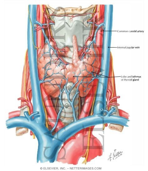
It supplies the thyroid gland. The neck is supplied by arteries other than the carotids. Bodytomy provides a labeled iliac artery diagram to help you understand the anatomy and function of the common iliac. Illustration showing arteries in the head noted at right are anterior cerebral artery middle cerebral artery internal carotid artery and common carotid artery labeled at left is posterior cerebral arteries in the neck the carotid arterial system lecturio from d3uigcfkiiww0g.cloudfront.net. Instant anatomy is a specialised web site for you to learn all about human anatomy of the body with diagrams, podcasts and revision questions. Left axillary a left vertebral a left carotid sinus right subclavian a. • external carotid artery terminates where it becomes the: Branches of arch of aorta. Arteries of the femoral neck. Terms in this set (32). Arteries of femoral head … The internal carotid artery (latin: Want to learn more about it?
Terms in this set (32). The left and right carotids, and the left and right vertebral arteries. It supplies the thyroid gland. Left axillary a left vertebral a left carotid sinus right subclavian a. Check interpret media out if you are looking for an illustrator.
Arteries of the neck - YouTube from i.ytimg.com From this trunk, several vessels arise, which go on to supply the neck. Arteria carotis interna) is located in the inner side of the neck in contrast to the external carotid artery. The first branch of the thyrocervical trunk is the inferior thyroid artery. The external carotid artery has eight named branches distributed to the head and neck. Instant anatomy is a specialised web site for you to learn all about human anatomy of the body with diagrams, podcasts and revision questions. Bodytomy provides a labeled iliac artery diagram to help you understand the anatomy and function of the common iliac. It supplies the thyroid gland. This entry was posted in anatomy, body parts, system and tagged anatomy of arteries, anatomy of artery, arteries, arteries anatomy, arteries chart, arteries diagram, arteries diagram with labels, arteries explained, artery.
Label the arteries of the neck in the ct angiogram.
The right and left subclavian arteries give rise to the thyrocervical trunk. Left axillary a left vertebral a left carotid sinus right subclavian a. Labeled diagram of the arteries of the head and neck. It is located on every side of the neck and is a large triangular space, with its apex pointed downwards and base pointed upwards and in front of… it's partially concealed by the posterior edge of the sternocleidomastoid. This diagram with labels depicts and explains the details of neck arteries. Is a delicate, subcutaneous muscle separating the skin from the deeper anterior muscles of the neck. The first branch of the thyrocervical trunk is the inferior thyroid artery. The principal arteries are the carotid and subclavian arteries. A blockage in one of the carotid arteries can be cleared either by endarterectomy or carotid angioplasty. The ascending aorta performs the function of supplying blood to the head, neck, and the arms, whereas. The coronary artery and the circumflex artery are responsible for delivering oxygenated blood to the heart and break it is also the biggest artery in the human body. All veins and arteries are in singular form, this will be easier for the test, i also removed left and right. It carries blood from the left ventricle to the coronary arteries.
The carotids reside beneath the skin on either side, and the pulse can be felt easily with your hand arteries in neck. Find the perfect neck label stock illustrations from getty images.


0 Komentar